Okayama University research: A novel 3D cell culture model sheds light on the mechanisms driving fibrosis in pancreatic cancer
OKAYAMA, Japan, Aug. 28, 2020 /PRNewswire/ -- In a recent study published in Biomaterials, researchers at Okayama University created a new 3D cell culture model of pancreatic cancer that closely mimics the "fibrotic" tissue characteristically observed in patients.
Pancreatic cancer is a lethal condition with a very poor prognosis—only ~9% of patients live to see another 5 years after diagnosis. A prime feature of pancreatic cancer is the presence of fibrotic tissue within the tumors. This fibrotic tissue is akin to the scarring that surrounds a wound. Fibrotic tissue entraps the cancer cells within it, making it difficult to therapeutically target these cells. Thus, understanding the mechanisms behind fibrotic tissue development is imperative for creating effective treatment strategies. Professor Mitsunobu R. Kano and Assistant Professor Hiroyoshi Y. Tanaka from Okayama University and colleagues have now created a three-dimensional (3D) cell culture model of pancreatic cancer in the laboratory which closely replicate the fibrotic nature of the tumors.
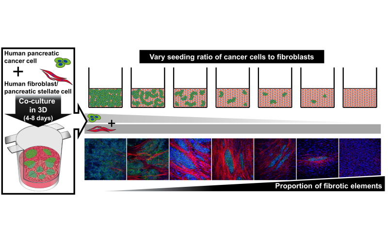
Fibrotic tissue develops when cancer cells and specialized cells called fibroblasts closely interact with each other. The patterns of fibrotic tissue seen in pancreatic cancer vary greatly from patient to patient. The researchers started by analyzing patient tumor samples and found that fibrotic tissue took up as little as 40% and as much as 80% of the space within tumors. For the 3D cell culture model to truly mimic the cancer, it would need to reflect this wide range in the amount of fibrotic tissue observed. To achieve this, the team tried seeding pancreatic cancer cells and fibroblasts at different ratios. Indeed, by trying various ratios, the team could create 3D pancreatic cancer tissues with any given amount of fibrotic tissue—most importantly within the clinically observed range.
The fibroblasts within these models were subsequently scrutinized to unravel cellular changes driving fibrotic tissue development. It was found that two proteins, namely, SMAD2/3 and YAP were the driving force behind such changes. These two proteins, however, did not act alone: the combined activity of SMAD2/3 and YAP were necessary for the fibroblasts to acquire the abnormal characteristics seen in tumor tissue. A host of cellular signaling systems were in place to enable the function of SMAD2/3 and YAP—some of these systems were common while others were unique to each protein.
Cell culture models of pancreatic cancer play an indispensable part in understanding the disease since they allow mechanistic analyses at a detail that would otherwise be difficult to achieve in studies using laboratory animals or clinical specimens. However, cell culture models to date generally failed to recreate the characteristic, densely fibrotic tissue observed in pancreatic cancer, much less the variability observed between patients. The 3D cell culture model of pancreatic cancer developed in this study overcomes these issues. The new model may enable researchers to understand the differences between tumors showing various degrees of fibrosis and potentially customize strategies to target them. "Our novel model will be useful in promoting the understanding of the complex mechanisms by which the fibrotic stroma develops and how it might be therapeutically targeted", conclude the researchers.
Background
Pancreatic cancer and fibrotic tissue: Pancreatic cancer is one of the most difficult to treat cancers. This is in large part due to the dense, fibrotic tissue present within the tumor.
Fibrosis is a biological process that occurs in damaged internal organs (such as the pancreas) when wound healing mechanisms go awry. Although fibrosis initiates as a process that protects a damaged organ, it sometimes also ends up creating an environment that promotes the growth of cancer cells. Thus, fibrotic tissue is closely associated with the presence and spread of pancreatic cancer. Fibrotic tissue also facilitates drug resistance thereby preventing the cancer cells from responding to any medication. Fibrotic tissue is therefore a huge barrier to understanding the complexities of pancreatic cancer and developing therapeutic strategies.
Link to figure
https://www.okayama-u.ac.jp/up_load_files/research_highlights/113_image_1.jpg
Caption
Pancreatic cancer cells (green areas) and fibroblasts (non-green areas) were mixed in specialized culture vessels to create a novel three-dimensional cell culture model of pancreatic cancer in the laboratory. The cells were mixed at various ratios to enable the tweaking of the amount of fibrosis, from almost no fibrotic tissue (bottom panel, leftmost image) to almost completely fibrotic tissue (bottom panel, 2nd image from the left). When cultured together with cancer cells, fibroblasts began to express a protein characteristic of cancer tissue (red) that was not observed when fibroblasts were cultured alone (bottom panel, left most image). The model thus successfully captures and allows the analysis of the interactions between cancer cells and fibroblasts that drive the formation of fibrotic tissue.
Reference
Hiroyoshi Y. Tanaka, Tsuyoshi Kurihara, Takuya Nakazawa, Michiya Matsusaki, Atsushi Masamune, Mitsunobu R. Kano. Heterotypic 3D pancreatic cancer model with tunable proportion of fibrotic elements. Biomaterials, 251, August 2020, 120077.
DOI : 10.1016/j.biomaterials.2020.120077
https://www.sciencedirect.com/science/article/pii/S0142961220303239
Reference (Okayama Univ. e-Bulletin): Professor KANO's team
OU-MRU Vol.62:3D tissue model offers insights into treating pancreatic cancer
Further information
Okayama University
1-1-1 Tsushima-naka , Kita-ku , Okayama 700-8530, Japan
Public Relations Division
E-mail: www-adm@adm.okayama-u.ac.jp
Website: http://www.okayama-u.ac.jp/index_e.html
Okayama Univ. e-Bulletin: http://www.okayama-u.ac.jp/user/kouhou/ebulletin/
Okayama University Medical Research Updates (OU-MRU)
Archive of all volumes:
https://www.okayama-u.ac.jp/eng/research/ou-mru.html
Vol.79:Novel blood-based markers to detect Alzheimer's disease
About Okayama University
Okayama University is one of the largest comprehensive universities in Japan with roots going back to the Medical Training Place sponsored by the Lord of Okayama and established in 1870. Now with 1,300 faculty and 13,000 students, the University offers courses in specialties ranging from medicine and pharmacy to humanities and physical sciences.
Okayama University is located in the heart of Japan approximately 3 hours west of Tokyo by Shinkansen.

Editor Details
-
Company:
- PR Newswire Europe
- Website:
